the instruments
The Crystal and the Beam
In 1935 a young researcher in Oxford took an X-ray photograph of an insulin crystal and saw that it was ordered enough to be solved. It took another thirty-four years to solve it.
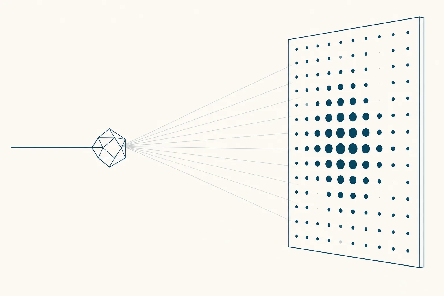
In the spring of 1935, in a damp basement room in the Oxford University Museum, a twenty-four-year-old researcher named Dorothy Crowfoot mounted a crystal of insulin in front of an X-ray tube and left it there overnight. When the plate was developed the following day it carried a pattern of spots — hundreds of them, arranged in a regular array, sharp enough to measure. She is said to have walked around Oxford for hours afterwards, too excited to work.
What the plate proved was not what insulin looked like. It proved that insulin had a definite shape at all: that every molecule in that crystal was folded the same way as every other, precisely enough to stack into a lattice and diffract as a unit. In 1935 that was a real question. Proteins were widely suspected of being ill-defined aggregates rather than discrete objects, and a photograph showing that one of them was as orderly as a mineral was a serious piece of evidence 1.
The structure itself took another thirty-four years.
Why a photograph is not a picture
A microscope works by collecting light scattered from an object and using a lens to reassemble it into an image. X-rays scatter from the electrons in a molecule in just the same way, and would produce a far finer image, except for one intractable fact: no lens exists that can bend them back together. The reassembly has to be done by calculation instead.
The calculation needs two numbers for every reflected spot — how strong it was, and where in its cycle the wave was when it arrived. The plate records the first faithfully. The second is destroyed in the act of measurement, because film and detectors register energy and are indifferent to phase. This is the phase problem, and it is not a technical shortcoming to be engineered away. It is a permanent hole in the middle of the data, and every method crystallography has ever developed is a strategy for filling it.
The strategy that worked for large molecules was to soak a heavy atom into the crystal — mercury, platinum, a lanthanide — and take the photographs again. A heavy atom scatters strongly and its own position can be worked out first, giving a reference against which everything else can be phased. Elegant in principle; in practice it meant persuading a fragile protein crystal to accept a foreign metal at one specific site without otherwise changing its packing, then repeating that with several different metals. Crystals cracked. Metals bound in the wrong places, or in too many. Some proteins never yielded a usable derivative at all.
What emerges from the calculation, once the phases have been supplied, is not a picture of atoms. It is a map of electron density — a three-dimensional contour surface showing where electrons are concentrated — and a chemist then has to build a molecular model into it, deciding which bump is a carbonyl and which is a side chain. How much that map constrains the decision depends on resolution. At coarse resolution the density is a smooth tube through which the chain evidently runs, and almost any plausible model can be forced into it. Only when the resolution is fine enough for individual side chains to lift out of the background does the map begin to arbitrate. A structure is therefore always an interpretation, and one of the enduring virtues of the technique is that the evidence behind the interpretation is preserved and can be re-examined by somebody else.
What the technique demanded of a sample
Behind the phase problem stood a more basic obstacle, and it is the reason crystallography advanced through the compounds it did rather than the ones anyone would have chosen.
The first protein ever to give a diffraction pattern illustrates the point exactly. In 1934, J. D. Bernal and Dorothy Crowfoot obtained X-ray photographs of crystalline pepsin, and the reason they succeeded where others had failed was almost comically mundane: previous workers had taken their crystals out of the liquid they grew in and let them dry, at which point the internal order collapsed and the pattern vanished. Bernal and Crowfoot sealed the crystal in a thin glass tube along with some of its mother liquor, so it stayed wet. Everything that followed in protein crystallography rests on that small procedural correction, and on the recognition behind it — that a protein crystal is more water than protein, and stops being a crystal the moment it stops being wet.
You cannot photograph what you cannot crystallise. A crystal is millions of copies of a molecule in identical orientation, and growing one requires material that is pure, abundant, and willing to settle into a single conformation. Any flexibility is fatal; a molecule with a floppy tail will either refuse to pack or will pack with that tail disordered, leaving a blur in the map where an answer should be. Crystallisation itself was, and largely remains, empirical — a matter of screening conditions and waiting, with no reliable theory of why one buffer works and a nearly identical one does not.
Then there was the arithmetic. Before electronic computers, the sums converting thousands of measured intensities into a map of electron density were performed by hand, with mechanical aids and printed tables, over months. Hodgkin's group worked through much of the penicillin analysis this way, and her Nobel lecture is candid about how much of the early work was labour rather than insight 3.
Penicillin, B12, and a prize
Penicillin was the proving ground. During the war its structure was genuinely contested: chemists had two candidate arrangements, and the argument mattered because nobody could plan a synthesis of a molecule whose shape was in dispute. Hodgkin's group settled it crystallographically, confirming the strained four-membered beta-lactam ring that most of the chemical establishment considered improbable. The structure was determined by 1945 and published in full afterwards, and it demonstrated something the field had not yet accepted — that crystallography could adjudicate a chemical question faster and more decisively than chemistry could.
Vitamin B12 was the harder case and the more astonishing result. It is a large, intricate molecule with a cobalt atom at its centre, and at the time of the analysis its constitution was almost entirely unknown; there was no chemical formula to check the answer against. The structure was published in 1956, worked out from the diffraction data with the help of some of the earliest electronic computing applied to the problem 2. The chemists had not merely failed to solve it — they were nowhere near solving it.
The 1964 Nobel Prize in Chemistry went to Hodgkin for determining by X-ray techniques the structures of important biochemical substances. She remains the only British woman to have received a Nobel Prize in any of the sciences.
1969, and the shape of a hormone
Insulin, meanwhile, had been running as a background project for three decades. It was a harder target than B12 by a wide margin, and it had to wait for heavy-atom derivatives that worked, for computers capable of the scale of calculation involved, and for a group large enough to sustain the effort. The structure of rhombohedral two-zinc insulin was published in 1969, with Hodgkin among a long list of authors reflecting how collective the work had become 4.
What emerged was not what the sequence alone would have suggested. Insulin in the crystal is not a lone chain but an assembly: six molecules arranged around two zinc ions, packed as three pairs. The surfaces the molecules use to hold each other turn out to be distinct from the surfaces that face outward, which raised an immediate and consequential question about which parts of the molecule are involved in binding a receptor and which are merely involved in storage. The follow-up review that Hodgkin wrote with Tom Blundell, Guy Dodson and Dan Mercola in 1972 works systematically through what the crystal structure implied for the chemistry and biology of the hormone, and reads now as a manifesto for a way of thinking that did not yet have a name 5.
The long shadow
That way of thinking is the real legacy, and it is easy to miss because it has become the ordinary condition of the field.
Before structures, modifying a biologically active molecule was a matter of chemical intuition and systematic variation. You changed something, made the compound, tested it, and inferred backwards from whether it still worked. The inference was weak, because a change that abolishes activity might have destroyed a binding contact or might merely have made the molecule fold differently, and there was no way to distinguish the two.
With a structure, the question becomes geometric. Which residues sit on the surface that contacts the partner? Which are buried and therefore structural? Where is there room for a substituent, and where would one collide? None of that makes design easy — a static picture of a crystal is a poor guide to a molecule in motion, and the history of the field is littered with compounds that fitted a structure beautifully and did nothing useful. But it converts guesswork into a reasoned argument that can be checked.
The idea took decades to become a discipline. Blundell went on to be among the people who built structure-based design into an industrial method; the antiviral programmes of the 1980s and 1990s, where a target enzyme's structure was solved and inhibitors were designed into its active site, were its first unambiguous commercial vindication. In the peptide field the same logic runs through every attempt to make a natural hormone longer-lived, more selective, or resistant to the enzymes that would otherwise clear it — each of those is an argument about a surface, and arguments about surfaces require a picture.
The measurement became routine in the way these stories always end. Solving a protein structure moved from a career to a thesis to, with synchrotron beamlines and automated crystallisation, a matter of weeks. A resource that began as a handful of coordinate sets holds hundreds of thousands of structures today, deposited as a matter of course. What took thirty-four years for insulin is now the least remarkable stage of a project — which is a strange fate for an instrument that once required a young researcher to walk around Oxford all afternoon because a plate of spots had come out sharp.